
Novel Spatial Technology May Improve the Understanding of Cancer Biology
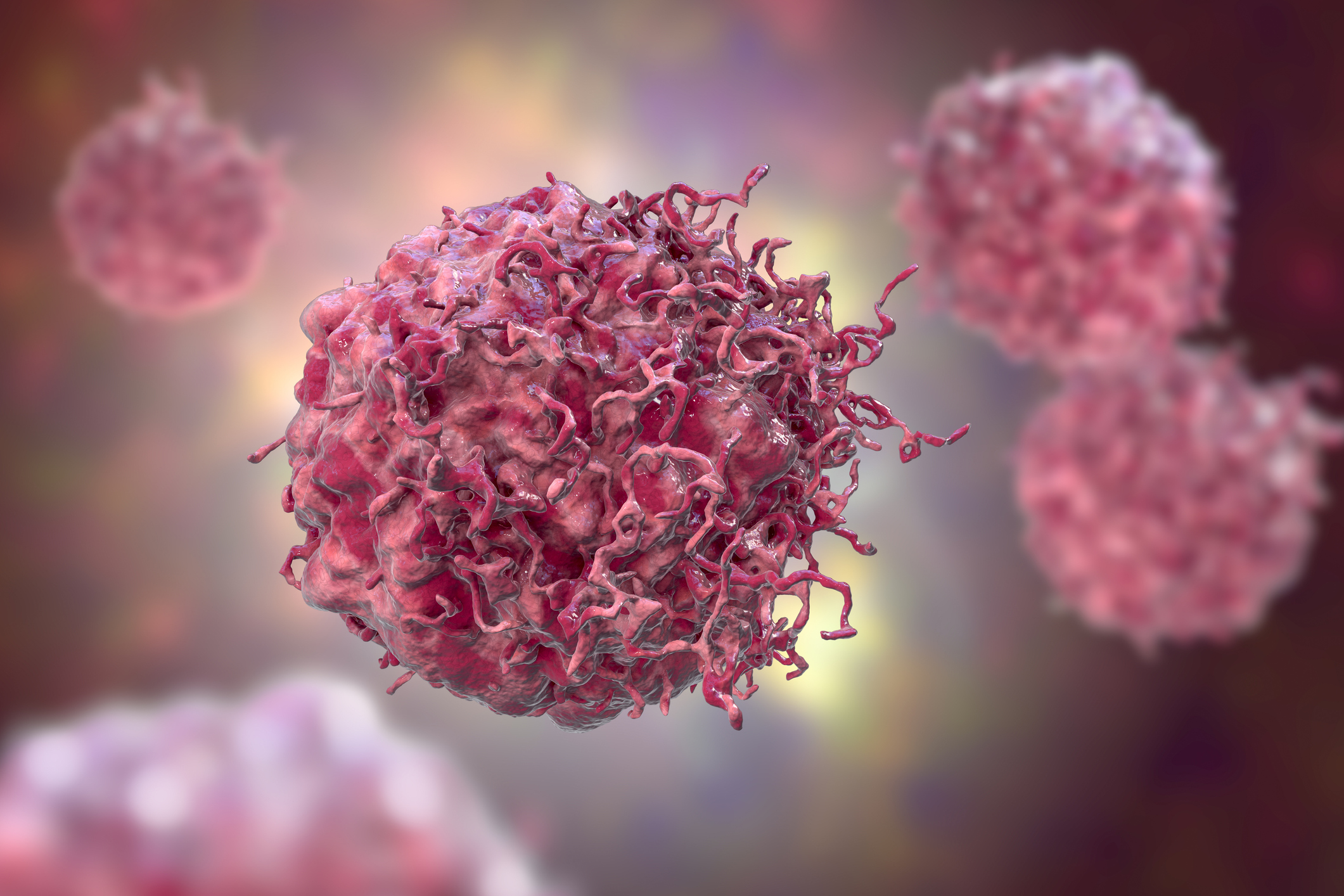
Northwestern Medicine scientists have developed a novel high-resolution spatial genomics platform that may enhance future research about how genome organization and genetic alterations contribute to the development of disease, including cancer, as detailed in a recent study published in Nature Methods.
“What is particularly exciting is that we can simultaneously profile 3D genome organization and chromatin accessibility while preserving the native spatial context of cells and tissues, and we can also detect genomic alterations such as copy-number changes and structural variations in cancer,” said co-corresponding author Feng Yue, PhD, the Duane and Susan Burnham Professor of Molecular Medicine and a professor of Biochemistry and Molecular Genetics and of Pathology.
Nearly all biological processes depend on the spatial organization of different types of cells within the tissue microenvironment, and the location and interactions of individual cells influence the function of these processes, which include protein localization, chromatin accessibility and histone modifications.
Recent advances in spatial “omics” techniques have enabled scientists to profile different molecular features, such as the transcriptome, proteome and epigenome while preserving their spatial context within tissues, but the techniques do not capture 3D genome folding.
Imaging-based approaches have also been used to study the genome organization and its relationship to biological processes such as immune response and disease progression. However, these approaches typically interrogate selected loci or regions in the genome, underscoring the need for complementary sequencing-based technologies that can map the entire genome.
To address this gap, Yue’s team developed Spatial-ATAC-Hi-C, a microfluidic-based spatial platform that can profile both 3D genome organization and chromatin accessibility within native tissue samples.
To validate their approach, the scientists applied Spatial-ATAC-Hi-C to adult mouse brain tissue and adult human brain tissue samples, revealing unique chromatin architecture features and gene regulatory programs in both neuronal and non-neuronal cell populations.
In glioblastoma and astrocytoma patient samples, Spatial-ATAC-Hi-C was also able to detect spatially resolved 3D genome alterations, in addition to genetic copy number variations and structure variations in certain regions of the tumors.
“We showed even within the same tumor, different regions can harbor distinct copy number alterations or structural variations,” said Yue, who is also director of the Center for Advanced Molecular Analysis. “Being able to detect these genomic differences while preserving their spatial context could help us better understand tumor heterogeneity.”
Overall, Spatial-ATAC-Hi-C provides a new technology for studying genome organization and gene regulation in cancer and developmental biology, according to Yue.
“This integrated approach provides a powerful tool to systematically explore the dynamic interplay between 3D chromatin structure, accessibility and spatiotemporal gene regulation within a complex biological context while preserving native spatial organization,” the authors wrote.
Ping Wang, PhD, research assistant professor of Biochemistry and Molecular Genetics; Juang Wang, PhD, a postdoctoral fellow in the Yue laboratory; Qixuan Wang, a student in the Driskill Graduate Program in Life Sciences (DGP); Mark Youngblood, MD, PhD, assistant professor of Neurological Surgery, were co-first authors of the study.
Rong Fan, PhD, Harold Hodgkinson Professor of Biomedical Engineering at Yale University, was co-corresponding author of the study.
Co-authors of the study include Alok Swaroop, ‘19 MD, ‘19 PhD, ’21 GME, instructor in the Department of Medicine, Division of Hematology and Oncology; Khizar Nandoliya, a medical student; Hinda Najem, PhD, a postdoctoral fellow in the Department of Neurological Surgery; Amy Heimberger, MD, PhD, the Jean Malnati Miller Professor of Brain Tumor Research and vice chair for research in the Department of Neurological Surgery; and Adam Sonabend, MD, the Etka Frimerman Professor of Neurosurgery.
Yue, Heimberger and Sonabend are members of the Robert H. Lurie Comprehensive Cancer Center of Northwestern University.
This work was supported by National Institute of Health grants R35GM124820, 1R01HG009906, and R01HG011207, T32 CA009560 and T32 CA070085; a Neurosurgery Research and Education Foundation Fellowship; the United States Department of Defense (CA230856); American Brain Tumor Association Basic Research Fellowship (fully supported by Tap Cancer Out); and the Society of Neurological Surgeons.
Related Stories
 Technology
TerraPower selects more suppliers for Natrium components
48 minutes ago
 Technology
UP to launch Indo
1 hour ago
 Technology
Bizzabo lays off dozens as Israeli event tech company restructures
1 hour ago
 Technology
Lagos’ tech valuations surge 12
2 hours ago
 Technology
Credo Technology Stock Tanks Despite Sales More Than Double
3 hours ago
 Technology
The IPO Window Is Closing. Here Are 8 Startups To Watch.
4 hours ago
 Technology
MIRAVA raises €4.2 million to build a new category in recreational boating (Sponsored)
5 hours ago
 Technology
Malanje certifies five space technology trainees as GGPEN strengthens local capacity
5 hours ago